Research · Building functional objects

Medical models and CT phantoms

Presurgical planning and training models printed directly from CT and MRI scans, with realistic appearance and feel and no manual segmentation.

The key idea. Medical scans are volumetric, but most printed anatomical models are opaque, rigid plastic shells traced from boundary surfaces, a process that can take a radiologist eight hours per model. OpenVCAD maps the scan volume directly onto a volumetric inkjet printer, so models keep the scan’s internal detail and can look and feel like real tissue, and those eight hours drop to seconds.

A hand holding a translucent printed block of a human chest, with orange ribs, the spine, and darker organs visible inside
A chest scan printed volumetrically on a full-color inkjet printer. Ribs, spine, and soft tissue keep the continuous gradations of the original scan.

Volumetric scans, volumetric prints

Most presurgical planning still happens on 2D slices of a scan. When models are 3D printed, they are usually made by segmentation: tracing the boundary of each tissue region into a separate surface mesh. That is slow, and it replaces the natural gradations between soft tissues with sharp boundaries. OpenVCAD instead imports the scan as a radiodensity field and, through design-intent compilation, maps it directly to printable properties, with no segmentation step.

Diagram comparing two workflows. Traditional segmentation goes from a DICOM image stack to segmented STL meshes, slicing, and printing. OpenVCAD goes from a DICOM image stack to a radiodensity attribute, a mapping to color, compilation, and a printed full-color head
Segmentation versus direct mapping. The traditional workflow (top) traces scan data into surface meshes, then slices and prints them. OpenVCAD (bottom) imports the scan as a radiodensity field, maps it to color, and compiles it straight to printer instructions, so the data stays volumetric from scan to print.
Two printed halves of a brain scan in blues and greens, with an oval yellow-green tumor in each half and a coin for scale
Printed brain with a tumor. A brain scan printed in two halves on a full-color inkjet printer. Scan values set color and opacity throughout, and the tumor shows in yellow-green.
The same model in OpenVCAD, previewed one layer at a time. Scan data: a 3D Slicer sample dataset.

One scan, different models

Because the scan data is never reduced to surfaces, the same scan can be translated for different purposes: color for visual inspection, or stiffness for a model that feels like real tissue in the hand and under surgical tools.
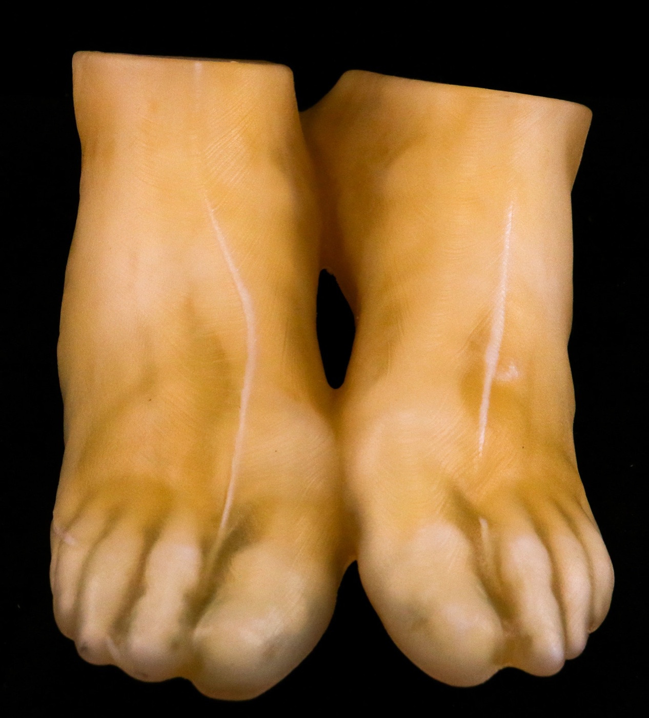
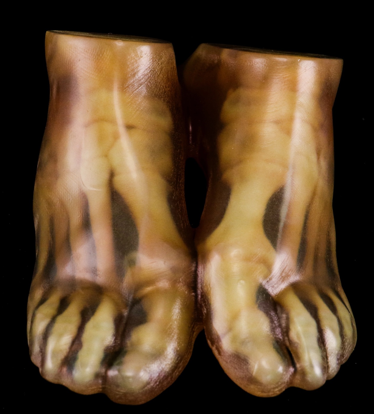
Printed pair of feet in pale tan, made of soft and rigid materials Printed pair of feet in translucent tan and brown with darker bones visible inside Visual model Mechanical model
One CT scan, two inkjet-printed models. For the visual model, radiodensity is mapped to color. For the mechanical model, it is mapped to stiffness, so bones print rigid while skin and heel stay soft. Drag the slider to compare.

CT phantoms

A phantom is an object made to be scanned. Phantoms are used to calibrate and check scanners and to train radiologists on standardized images. Traditional phantoms are usually assembled by hand from many parts, each with its own radiodensity, which limits how much anatomy they can represent. Here, a CT scan of a real person’s feet was imported into OpenVCAD with radiodensity as the design intent. A material mapping we developed converted it into mixtures of a radiopaque inkjet resin (RadioMatrix) and a conventional non-radiopaque resin, and the printed phantom was scanned again. Because phantoms like this come straight from scan data, they can represent a much wider range of realistic anatomy, including atypical conditions.
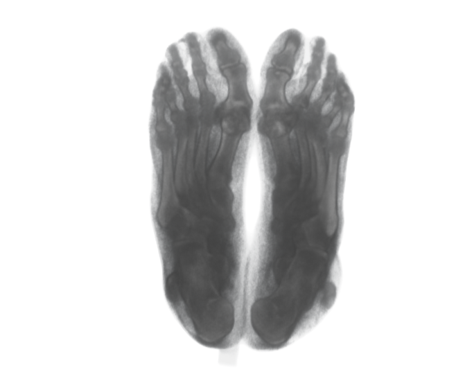
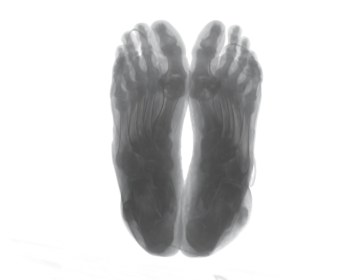
CT image of the printed phantom feet seen from below, closely matching the original CT image of a person's feet seen from below Original CT CT of the print
The original scan and a scan of the printed phantom. The print reproduces the spatial variation of the patient’s CT. Drag the slider to compare.
A dark, glossy printed phantom shaped like a pair of feet
The printed phantom. Its dark color comes from the resins; what matters is how it appears in a CT scan.

Vascular models

The same tools design vascular networks at realistic sizes. Each training model holds more than 20 million voxels of multi-material assignments that mimic how real tissue responds to tools: graded soft and hard regions, such as a stiffer sheath around each vein, and liquid printed inside the vessels by the inkjet printer. The models can be cut and bleed, and they let medical students practice drawing blood, including the way a real vein rolls away from a needle. This is a collaboration with Joseph Passarello, a master’s student I mentor, funded by a grant from the Defense Health Agency. Every model is generated by customizable OpenVCAD scripts.

A clear printed block containing a branching network of red vessels
A printed vascular network inside a clear block.
Drawing blood from an inkjet-printed training model designed in OpenVCAD.
Cutting a vessel, which bleeds the liquid printed inside it.

Publications

  1. Design-Intent Compilation for Heterogeneous Fabrication
    Design-Intent Compilation for Heterogeneous Fabrication
    Charles Wade, Devon Beck, and Robert MacCurdy
    2026
    In submission · arXiv preprint